Anterior Fontanelle Closure Radiology

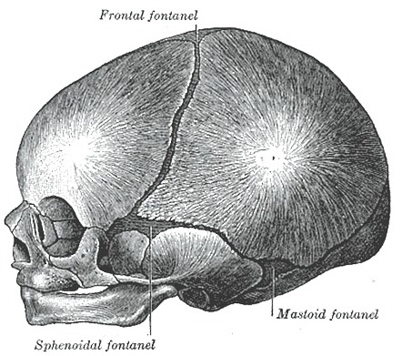
At birth an infant has six fontanels.
Anterior fontanelle closure radiology. Roentgenographically detectable bulging of the anterior fontanelle did not result from transient increases in intracranial pressure e g. The anterior or frontal fontanelle is the diamond shaped soft membranous gap at the junction of the coronal and sagittal sutures. The fontanelle at the top of the head anterior fontanelle most often closes between 7 to 19 months.
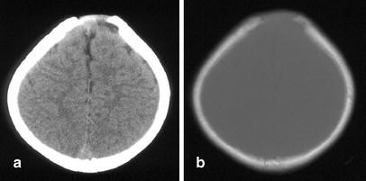
10 1055 b 0034 87890 sutures and fontanelles the width of the sutures is highly variable in neonates. Of the anterior fontanelle. A lateral view of skull soft tissue technique 45 kv shows mass located beneath normal layer of subcutaneous fat at anterior angle of fontanelle.
Widened sutures as a symptom of defective ossification in this instance no signs of increased icp are present. The fontanelle in the back of the head posterior fontanelle most often closes by the time an infant is 1 to 2 months old. Plain film ultrasound us and computed tomography ct can be used for assessment of sutural patency.
In chimpanzees the anterior fontanelle is fully closed by 3 months of age. The diagnosis of an abnormal fontanel requires an understanding of the wide variation of normal. The skull shape then undergoes characteristic changes depending on which suture s close early.
The coronal suture is the first to manifest widening in response to increased intracranial pressure icp. In contrast apes fuse the fontanelles soon after birth. Bony margins of fontanelle are nor mal.
The anterior fontanel is the largest and most important for. The average size of the anterior fontanel is 2 1 cm and the median time of clo. Suture widening is often associated with numerous wormian bones and a persistent fontanelle.


